
OptoTree’s software and biomarkers are ideally suited for analyzing images from the latest OCT-A retinal scanners from companies such as Carl Zeiss AG, Topcon Corporation, Heidelberg Engineering GmbH, Optopol Technology – REVO OCT, and OptoVue, Inc. These OCT-A scanners non-invasively generate contrasted image retinal micro-vascular structures and blood flow in three dimensions without, as is the case for fluorescein angiography, the need of a contrast agent to be injected into the eye. OCT-A scans can also be conducted in seconds versus 15 – 30 minutes, which is the case for fluorescein angiography, the current 2D standard for diagnosing and assessing diabetic retinopathy (DR). OptoTree’s automated and quantitative evaluation is also expected to provide more consistent assessments than of individual retina specialists, which today perform qualitative (as opposed to quantitative) assessments with variable results based on each specialist’s skill and experience. Notwithstanding the potential variability in qualitative assessments performed by different specialists, the number of specialists capable of interpreting fluorescein angiographs is grossly inadequate to review the number of eyes needing this examination.
OptoTree expects its Vasculomics®-derived DR biomarkers and analytical services, all delivered through the cloud, will provide high value-added applications for these OCT-A machines. Management believes these applications will quickly give clinical retina practitioners much more sensitive, precise, reliable, and actionable quantitative information enabling earlier diagnosis of disease presence and stage, and more precise and tightly controlled treatment regimens and monitoring. The Company expects OCT-A scanner manufacturers will benefit from the OptoTree Vasculomics® Platform since these tools will end up driving demand for OCT-A scanners.
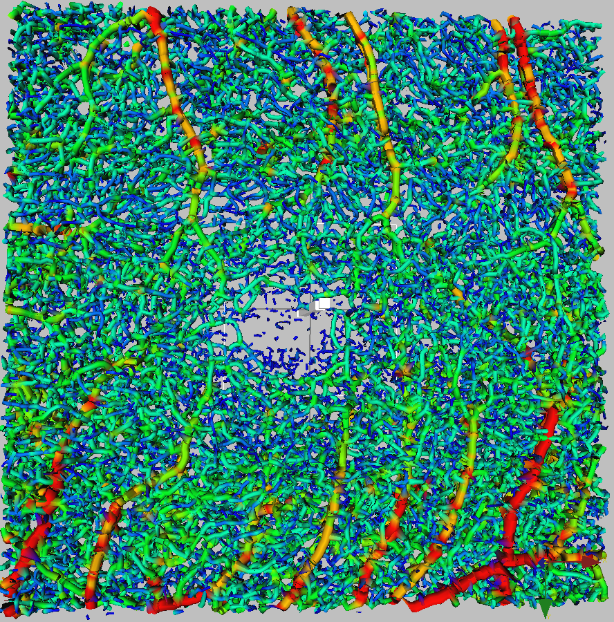
To see a sample image that gets closer to the detail available with our Vasculomics® technology, click the following thumbnail image:

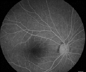
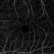
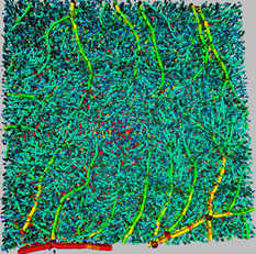
Retina Vasculomics® DR Examples
Vertical Columns – DR Stages, Horizontal Rows – Retina Analytical Modes
| Healthy, No Diabetes | Diabetes, Mild DR | Diabetes, Severe DR | |
| AngioVasc® Mode |  | 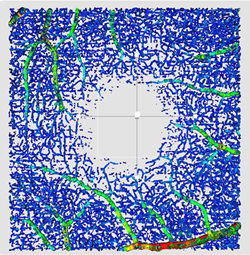 | 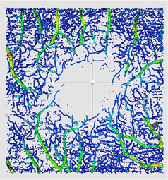 |
| AngioPerf® Mode |  | 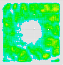 | 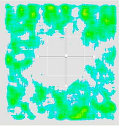 |
Vasculomics® Overview
Bio-Tree has spent over a decade developing and using the Vasculomics® technology and software platform, initially used for pre-clinical oncology drug discovery and toxicology projects with large pharmaceutical companies. This same platform and the extensive 3-dimensional computational micro-vascular imaging analysis know-how has been applied and adapted to analyze retina OCT-A datasets. Cancer and retinal disease express themselves similarly in tumor vessels and retina vessels by the formation of neo-vasculature that is chaotic, tortuous and curvy. Retina vessels have similar disease behavior and response to therapy as cancerous tumor vessels. Both tumor angiogenesis and retina vessel angiogenesis are being treated with the same anti-angiogenic VEGF inhibitors – e.g. Avastin, Tarceva, Lucentis, Eylea, etc. The Vasculomics® technology platform has been used successfully in detecting discovery stage tumor angiogenesis and its response to therapy. In the retina, it is being used in conjunction with OCT Angiography scanners to detect DR caused retina vascular changes and their response to therapy. These retina vascular changes are similar to the processes leading to tumor angiogenesis, including neo-vascular proliferation.